The Gastrolab Image Gallery
COLLAGES: Oesophagus

Collage:
Larynx

Collage:
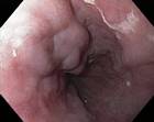
A Normal Oesophagus and Cardia

Collage:
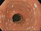
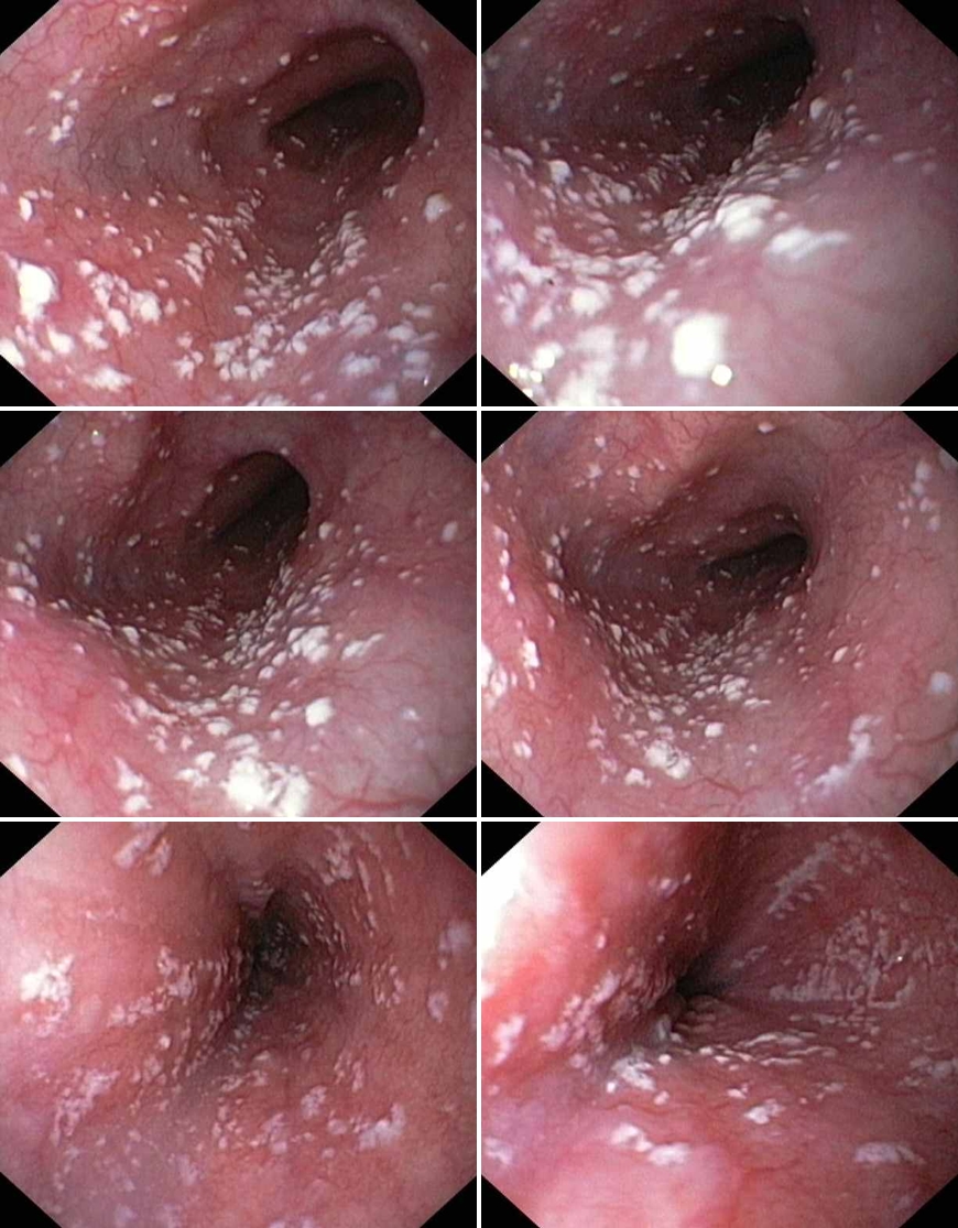
Glycogenic Acanthosis
Collage:
Oesophageal Inlet Patch

Collage:
Oesophageal Diverticulum (in the middle third)

Collage:
Oesophageal Diverticulum (in the middle third)

Collage:
Oesophageal Diverticulum (in the middle third)

Collage:
Oesophageal Diverticulum (in the middle third)

Collage:
Candida-oesophagitis
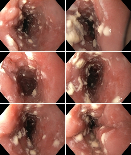
Collage:
Candica-oesophagitis
Collage:
Candica-oesophagitis

Collage:
Oesophageal varices in portal hypertension

Collage:
Oesophageal varices

Collage:
Oesophageal pseudovarices

Collage:
Corrosive oesophagitis, caused by NSAIDs

Collage:
Oesophagitis caused by vomiting

Collage:
Oesophagitis caused by vomiting

Collage:
Oesophagitis caused by vomiting

Collage:
Oesophageal drug injury (Alendronate)

Collage:
Oesophageal Web or Ring

Collage: Oesophageal Lesion Caused by a Sharp Chicken Bone

Collage:
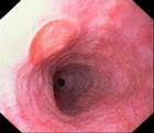
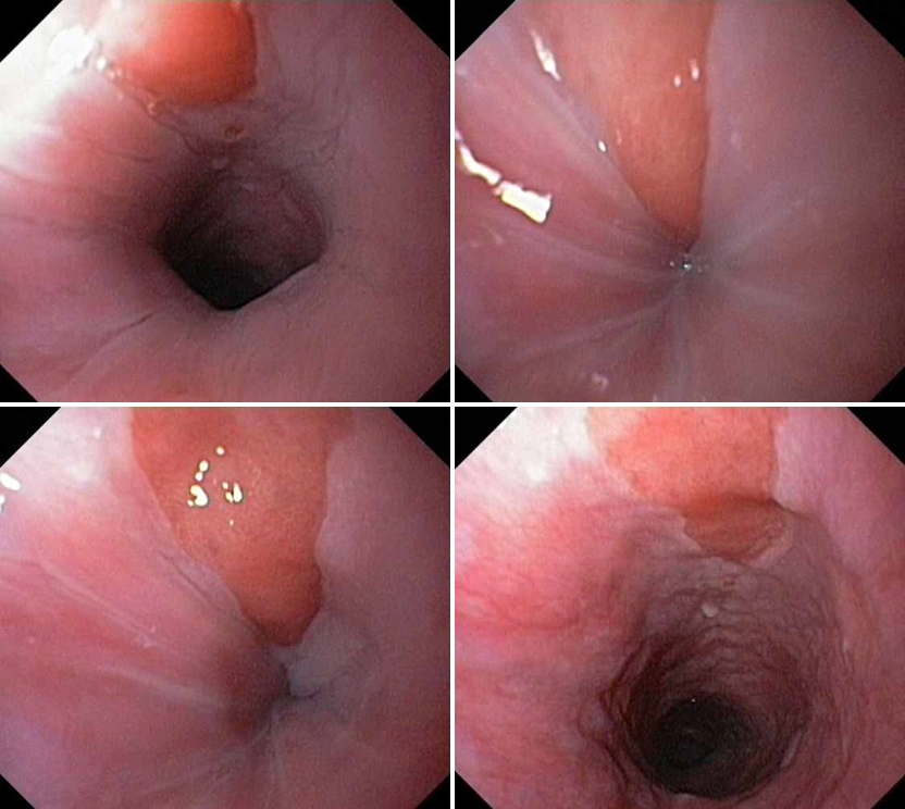
Barretts oesophagus
Collage:
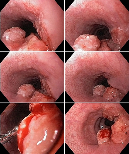
Oesophageal squamocellular cancer

Collage:
Oesophageal squamocellular cancer
Collage:
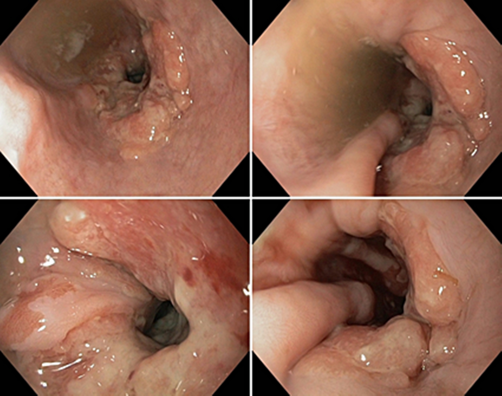
Oesophageal adenocarcinoma

Collage:
Oesophageal tuberculosis (The last image after anti-tbc treatment)