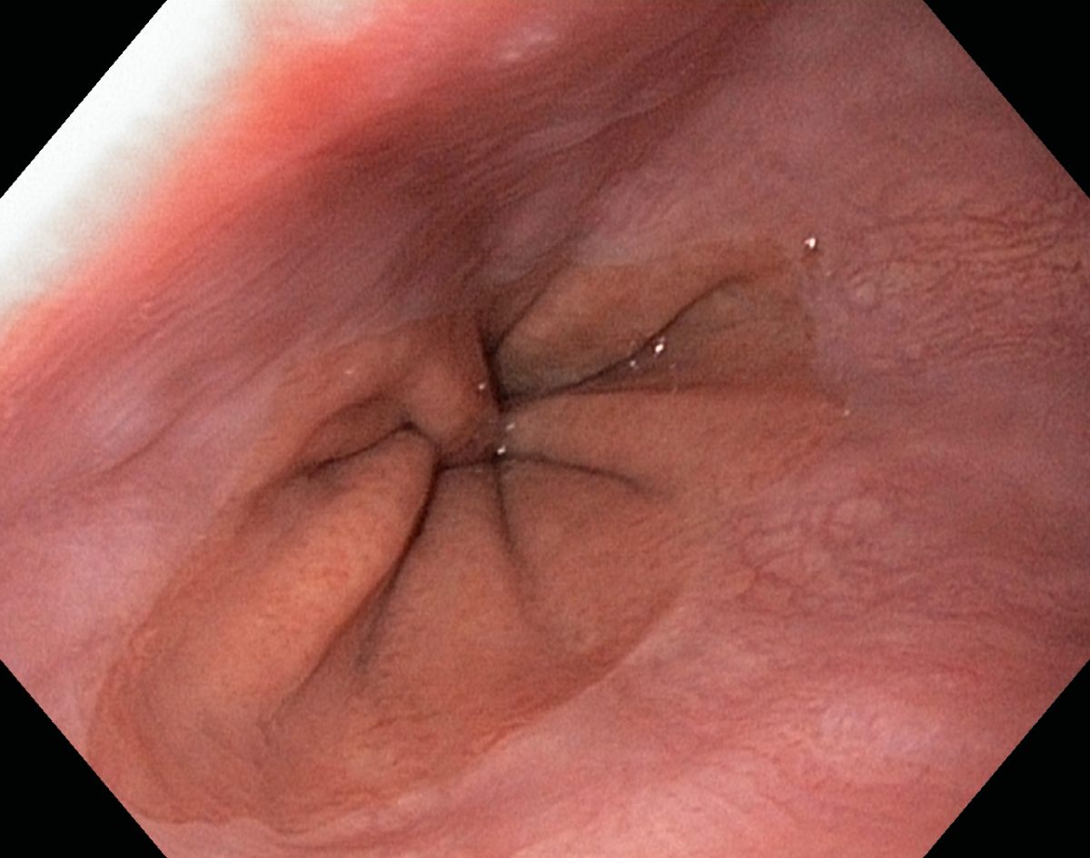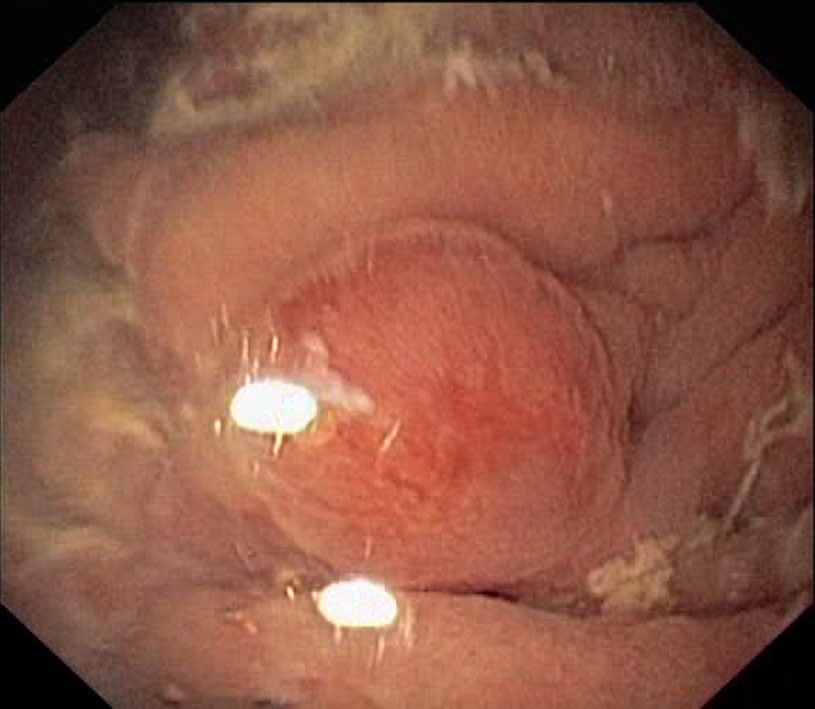
A normal oesophagus

A normal oesophagus

A normal oesophagus

Oesophageal varices

Oesophageal varices

Oesophageal varices

A normal cardia

Severe chronic gastritis caused by an Helicobacter-infection

Severe chronic gastritis caused by an Helicobacter-infection

Severe chronic gastritis caused by an Helicobacter-infection

Intestinal metaplasia on the gastric corpus in a patient with atrophic gastritis and pernicious anaemia

A normal descending part of the duodenum

A normal descending part of the duodenum

A normal descending part of the duodenum

Colon cancer (adenocarcinoma) in the ascending colon

Colon cancer (adenocarcinoma) in the ascending colon

Colon cancer (adenocarcinoma) in the ascending colon

Colon cancer (adenocarcinoma) in the ascending colon

Inflammatory polyposis caused by a chronic ulcerative colitis

Inflammatory polyposis caused by a chronic ulcerative colitis

Inflammatory polyposis caused by a chronic ulcerative colitis

Inflammatory polyposis caused by a chronic ulcerative colitis

Tubular adenoma in the descending colon

Tubular adenoma in the descending colon

Tubular adenoma in the descending colon

Tubular adenoma in the descending colon - endoscopic polypectomy

Scar after polypectomy (tubular adenoma in the descending colon)

Sigmoid diverticulas

Rectal venectasiaes (dilated veins)

Rectal venectasiaes (dilated veins)

Rectal venectasiaes (dilated veins)

A rectal tubular adenoma

A laterally spreading sessile tubular adenoma with a small cancer (adenocarcinoma) in the rectum

A laterally spreading sessile tubular adenoma with a small cancer (adenocarcinoma) in the rectum

A laterally spreading sessile tubular adenoma with a small cancer (adenocarcinoma) in the rectum

A laterally spreading sessile tubular adenoma with a small cancer (adenocarcinoma) in the rectum

These rectal small round mucosal spots seen in a colonoskopy - what are they? Any suggestions - please send an email (hansbjorknas@gmail.com).

These rectal small round mucosal spots seen in a colonoskopy - what are they? Any suggestions - please send an email (hansbjorknas@gmail.com).

These rectal small round mucosal spots seen in a colonoskopy - what are they? Any suggestions - please send an email (hansbjorknas@gmail.com).

These rectal small round mucosal spots seen in a colonoskopy - what are they? Any suggestions - please send an email (hansbjorknas@gmail.com).

Internal haemorrhoids

Oesophageal squamocellular cancer

Oesophageal squamocellular cancer

Oesophageal squamocellular cancer

Oesophageal squamocellular cancer

Cardia

A small carcinoid tumour in the gastric body in a patient with atrophic gastritis

A small carcinoid tumour in the gastric body in a patient with atrophic gastritis

A small carcinoid tumour in the gastric body in a patient with atrophic gastritis

GAVE, Gastric Antral Vascular Ectasia

GAVE, Gastric Antral Vascular Ectasia

GAVE, Gastric Antral Vascular Ectasia

GAVE, Gastric Antral Vascular Ectasia

Antrum and Pylorus

A normal duodenal bulb

A normal duodenal bulb

Duodenal ulcer disease

Duodenal ulcer disease

Duodenal ulcer disease

Tubular adenoma adjacent to the papilla Vater in a patient with FAP

Tubular adenoma adjacent to the papilla Vater in a patient with FAP

Tubular adenoma adjacent to the papilla Vater in a patient with FAP

Tubular adenoma adjacent to the papilla Vater in a patient with FAP

Tubular adenoma adjacent to the papilla Vater in a patient with FAP

Duodenal mucosa in Coeliac Disease

Duodenal mucosa in Coeliac Disease

Duodenal mucosa in Coeliac Disease

Duodenal mucosa in Coeliac Disease

Crohns disease in the ileum

Crohns disease in the ileum

Crohns disease in the ileum

Crohns disease in the ileum

Crohns disease in the ileum

Crohns disease in the ileum

Valvula Bauhini, the Ileocaecal Valve (to the left in this image)

Endoscopic polypectomy (tubular adenoma in the colon

Endoscopic polypectomy

Scar after polypectomy

Scar after polypectomy and the removed polyp to the right

Endoscopic polypectomy, tubular adenoma in the colon

Endoscopic polypectomy, tubular adenoma in the colon

Endoscopic polypectomy, tubular adenoma in the colon

Endoscopic polypectomy, tubular adenoma in the colon

Endoscopic polypectomy, tubular adenoma in the colon

Endoscopic polypectomy, tubular adenoma in the colon - smoke and scar

Faecesfilled diverticulum in the sigmoid colon

Sigmoid diverticulas
Click on the picture to get a magnification
Barretts oesophagus
Click on the picture to get a magnification
A short Barretts oesophagus
Click on the picture to get a magnification
Cameron-ulcus
Click on the picture to get a magnification
Cameron-ulcus
Click on the picture to get a magnification
Small hyperplastic polyps in the gastric fundus
Click on the picture to get a magnification
Occult mucosal bleeding (due to the use of NSAIDs) in the gastric antrum
Click on the picture to get a magnification
A normal descending part of the duodenum
Click on the picture to get a magnification
Papilla of Vater in the descending part of the duodenum
Click on the picture to get a magnification
Terminal ileum with the villi clearly visible
Click on the picture to get a magnification
Ileocaecal valve (Valve Bauhini) and caecum
Click on the picture to get a magnification
Ileocaecal valve
Click on the picture to get a magnification
Lipomatous ileocaecal valve
Click on the picture to get a magnification
Appendix aperture in the caecum
Click on the picture to get a magnification
A small caecal angiodysplasia
Click on the picture to get a magnification
Tubular adenoma in the transverse colon
Click on the picture to get a magnification
Diverticulosis in the sigmoid colon
Click on the picture to get a magnification
Diverticulum in the sigmoid colon
Click on the picture to get a magnification
Internal haemorrhoids (seen with the endoscope in an inverted position in the rectum(
Click on the picture to get a magnification
Hiatal hernia seen from below with the endoscope in the stomach in an inverted position
Click on the picture to get a magnification
Hiatal hernia seen from below with the endoscope in the stomach in an inverted position
Click on the picture to get a magnification
Hiatal hernia seen from below with the endoscope in the stomach in an inverted position
Click on the picture to get a magnification
Hiatal hernia seen from above, inside view
Click on the picture to get a magnification
A pediatric gastroscope with a diameter of just over 5 mm
Click on the picture to get a magnification
Larynx seen with a pediatric gastroscope with a diameter of just over 5 mm
Click on the picture to get a magnification
The gastric body seen with a pediatric gastroscope with a diameter of just over 5 mm
Click on the picture to get a magnification
The descending duodenum seen with a pediatric gastroscope with a diameter of just over 5 mm
Click on the picture to get a magnification
Hiatal hernia seen from above, inside view
Click on the picture to get a magnification
Gastric cancer (adenocarcinoma of the intestinal type) causing gastric retention
Click on the picture to get a magnification
Duodenal diverticulum (in the descending part of the duodenum)
Click on the picture to get a magnification
Duodenal diverticulum (in the descending part of the duodenum)
Click on the picture to get a magnification
Duodenal diverticulum (in the descending part of the duodenum)
Click on the picture to get a magnification
Lymphoid hyperplasia in the terminal ileum
Click on the picture to get a magnification
Lymphoid hyperplasia in the terminal ileum
Click on the picture to get a magnification
Crohns disease in a neoterminal ileum
Click on the picture to get a magnification
(Pseudo-)Melanosis coli caused by an exsessive use of laxatives
Click on the picture to get a magnification
(Pseudo-)Melanosis coli caused by an exsessive use of laxatives
Click on the picture to get a magnification
(Pseudo-)Melanosis coli caused by an exsessive use of laxatives
Click on the picture to get a magnification
A typical colon cancer (adenocarcinoma) in the ascending colon
Click on the picture to get a magnification
A typical colon cancer (adenocarcinoma) in the ascending colon
Click on the picture to get a magnification
A typical colon cancer (adenocarcinoma) in the ascending colon
Click on the picture to get a magnification
Endoscopic polypectomy (a tubular adenoma in the sigmoid colon)
Click on the picture to get a magnification
Endoscopic polypectomy (a tubular adenoma in the sigmoid colon)
Click on the picture to get a magnification
A circular ulcer in a neoterminal ileum caused by Crohns disease
Click on the picture to get a magnification
A circular ulcer in a neoterminal ileum caused by Crohns disease
Click on the picture to get a magnification
A distal ulcerative colitis
Click on the picture to get a magnification
A distal ulcerative colitis
Click on the picture to get a magnification
A distal ulcerative colitis
Click on the picture to get a magnification
A typical ulcerative proctitis
Click on the picture to get a magnification
A typical ulcerative proctitis
Click on the picture to get a magnification
A typical ulcerative proctitis
Click on the picture to get a magnification
A small cancer (adenocarcinoma) in the ascending colon
Click on the picture to get a magnification
A small cancer (adenocarcinoma) in the ascending colon
Click on the picture to get a magnification
Oesophageal web or ring
Click on the picture to get a magnification
A short Barretts oesophagus
Click on the picture to get a magnification
A short Barretts oesophagus
Click on the picture to get a magnification
Hiatal Hernia seen with the endoscope in an inverted position
Click on the picture to get a magnification
GAVE (Gastric Antral Vascular Ectasia)
Click on the picture to get a magnification
GAVE (Gastric Antral Vascular Ectasia)
Click on the picture to get a magnification
Duodenal mucosa with the (lipidfilled?) villi clearly visible
Click on the picture to get a magnification
Lymphoid hyperplasia in the terminal ileum
Click on the picture to get a magnification
A typical ulcerative colitis
Click on the picture to get a magnification
A typical ulcerative colitis
Click on the picture to get a magnification
A typical ulcerative colitis
Click on the picture to get a magnification
Active ulcerative colitis in the distal colon
Click on the picture to get a magnification
Active ulcerative colitis in the distal colon
Click on the picture to get a magnification
Active ulcerative colitis in the distal colon
Click on the picture to get a magnification
Active ulcerative colitis in the distal colon
Click on the picture to get a magnification
A normal larynx
Click on the picture to get a magnification
A normal oesophagus
Click on the picture to get a magnification
A normal oesophagus
Click on the picture to get a magnification
Candica oesophagitis
Click on the picture to get a magnification
Oesophageal varices caused by NASH with cirrhosis
Click on the picture to get a magnification
Oesophageal varices caused by NASH with cirrhosis
Click on the picture to get a magnification
Hyperplastic polyps in the gastric fundus
Click on the picture to get a magnification
A normal gastric antrum and pylorus
Click on the picture to get a magnification
A normal gastric antrum and pylorus
Click on the picture to get a magnification
A small xanthelams (xanthoma) in the gastric antrum
Click on the picture to get a magnification
A small xanthelams (xanthoma) in the gastric antrum
Click on the picture to get a magnification
Prepyloric benign gastric ulcer
Click on the picture to get a magnification
A benign prepyloric ulcer with a Forrest II lesion
Click on the picture to get a magnification
A benign prepyloric ulcer with a Forrest II lesion
Click on the picture to get a magnification
A benign prepyloric ulcer with a Forrest II lesion
Click on the picture to get a magnification
A normal duodenal bulb
Click on the picture to get a magnification
A huge duodenal ulcer
Click on the picture to get a magnification
A huge duodenal ulcer
Click on the picture to get a magnification
Caecum and the appendix aperture
Click on the picture to get a magnification
Ascending colon and the ileocaecal valve
Click on the picture to get a magnification
Caecal cancer (Adenocarcinoma)
Click on the picture to get a magnification
Caecal cancer (Adenocarcinoma)
Click on the picture to get a magnification
Caecal cancer (Adenocarcinoma)
Click on the picture to get a magnification
Hepatic flexure with the liver visible through the colonic wall
Click on the picture to get a magnification
Colon transversum, the transverse part of the large bowel, a normal endoscopic finding
Click on the picture to get a magnification
Diverticulas in the sigmoid colon
Click on the picture to get a magnification
A distal ulcerative colitis - the border between normal mucosa and colitis in the sigmoid colon
Click on the picture to get a magnification
A tubular adenoma in the sigmoid colon
Click on the picture to get a magnification
A small cancer (adenocarcinoma) in the sigmoid colon
Click on the picture to get a magnification
Scars after polypectomy in the colon
Click on the picture to get a magnification
A large bleeding rectal cancer (adenocarcinoma)
Click on the picture to get a magnification
A large rectal cancer (adenocarcinoma)
Click on the picture to get a magnification
A large rectal cancer (adenocarcinoma)
Click on the picture to get a magnification
A large rectal cancer (adenocarcinoma)
Click on the picture to get a magnification
A large rectal cancer (adenocarcinoma)
Click on the picture to get a magnification
A large rectal cancer (adenocarcinoma)
Click on the picture to get a magnification
Oesophageal pseudovarices
Click on the picture to get a magnification
Hiatal hernia and a short Barretts oesophagus
Click on the picture to get a magnification
A bleeding cardia cancer (adenocarcinoma)
Click on the picture to get a magnification
Cardia cancer (adenocarcinoma) seen from below with the endoscope in an inverted position
Click on the picture to get a magnification
Hyperplastic polyps in the gastric body
Click on the picture to get a magnification
Gastric retention and a gastric cancer (adenocarcinoma of the intestinal type) in the gastric body
Click on the picture to get a magnification
Gastric retention caused by diabetic gastroparesis
Click on the picture to get a magnification
Large inflammatory polyp in the gastric antrum
Click on the picture to get a magnification
Erosive duodenitis
Click on the picture to get a magnification
Duodenal ulcer
Click on the picture to get a magnification
Duodenal lipoma
Click on the picture to get a magnification
An Asacol(TM)tablet in the caecum
Click on the picture to get a magnification
Scars in the colon caused by ulcerative colitis
Click on the picture to get a magnification
Colon cancer (adenocarcinoma) in the ascending colon
Click on the picture to get a magnification
Colon cancer (adenocarcinoma) in the ascending colon
Click on the picture to get a magnification
Colon cancer (adenocarcinoma) at the splenic flexure
Click on the picture to get a magnification
Tubular adenoma in the sigmoid colon, polypectomysnare
Click on the picture to get a magnification
A patchy ulcerative colitis in the sigmoid colon
Click on the picture to get a magnification
A large malignant rectal polyp (tubulovillous adenoma and adenocarcinoma)
Click on the picture to get a magnification
A large ulcerating and necrotic rectal cancer (adenocarcinoma)
Click on the picture to get a magnification
A benign rectal tubular adenoma
You might also be interested in:
 A normal oesophagus
 |
 Hiatal Hernia from Below
 |
 A Normal Antrum and Pylorus
 |
 A Normal Descending Duodenum
 |
 A Normal Caecum
 |

